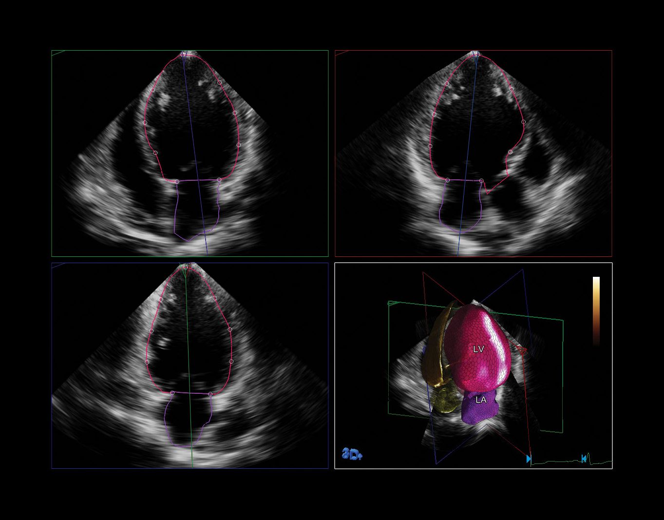
Cardiovascular diseases are the number one cause of death globally [1]. To deliver an accurate diagnosis, clinicians often use 2D ultrasound to measure the heart’s left chambers. But these exams can underestimate results because of image foreshortening and incomplete data. While 3D techniques can overcome this issue, many are cumbersome and tedious. Philips HeartModel* is a 3D tool that enables fast exam times, seamless workflow and broad applicability. It automatically detects, segments and quantifies the left ventricle (LV) and left atrium (LA) from live 3D images.
Chronic disease populations are growing, requiring the need to effectively and efficiently collect and analyze heart measurements in order to manage and better plan therapy for more patients in less time. HeartModel is an intuitive 3D tool that helps simplify this complicated exam and make it accessible for everyday clinical practice. It helps clinicians to quickly, easily and confidently assess disease states, determine treatment.
In only a few seconds, the application can quickly measure reproducible ejection fraction (EF). HeartModel offers easy and fast 3D images of the heart and is one of the only tools that can simultaneously calculate left ventricle (LV) and left atrium (LA) volumes from a single volume loop, equal to the period of time it takes for one heartbeat.

![HeartModel provides time savings of up to 82% compared to 3D manual measurements, with no user interaction necessary [2].](https://images.philips.com/is/image/philipsconsumer/6e0d86210d524bc6a875b22100f02ffd?extend=0,0,0,0&wid=450&hei=450&fit=stretch,1&$jpglarge$)
HeartModel is an example of how Philips is driving innovation in the cardiology space to help clinicians make decisions early, quickly and confidently. It is part of a suite of tools and technologies available on Philips’ EPIQ CV ultrasound systems.
This technology is designed to help address some of the most critical strains on overburdened hospitals and healthcare systems challenged to provide higher quality care at a lower cost.
